


Level Up- When The Meniscus Shock Absorber Hurts
LEVELUP INSIDERS December 14, 2017 Nirav Pandya 0
The dreaded swollen knee. As fall sports are in full swing we unfortunately see a large number of athletes who tumble to the ground; grabbing a knee in severe pain. Swelling in the knee indicates a joint which is aggravated and may have multiple forms of internal damage including a kneecap dislocation, ligament tear, or fracture. Another common diagnosis is a tear of the meniscus.
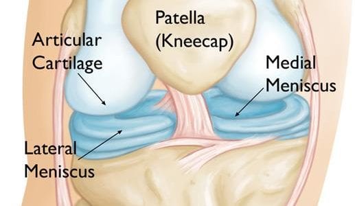
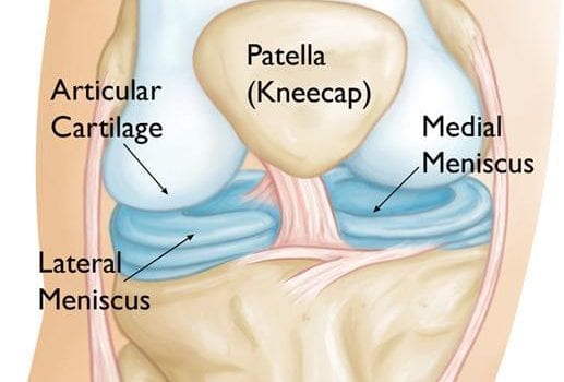
The meniscus is a shock absorber located between the femur (thigh) and tibia (shin) bone. The meniscus also acts as a secondary stabilizer. There are two menisci in the knee: one medial (inside) and one lateral (outside). When the meniscus tears, particularly in a young athlete, we are inclined to treat the injury surgically due to increased activity demands, a greater healing potential and because the injury is generally acute. Meaning it occurs from a traumatic event rather than wear and tear from years of activity.
After examining your knee and obtaining x-rays, a doctor will typically order a MRI to look for a meniscus tear and any additional damage to ligaments, cartilage, and tendons. If a meniscus tear is found, it can occur in different patterns: bucket handle, horizontal, longitudinal, flap, free edge fraying, radial tears and complex combination tears. If the tear warrants surgery, there are multiple options.
The surgeon will typically perform the surgery with an arthroscope, a small camera inserted into the knee. They will then assess the tear to determine its size and location. This is important as there are two main ways you can treat a meniscus tear: remove the torn portion or repair it. The decision to repair (sew together) versus removing the torn piece depends largely on the blood supply to the meniscus. The meniscus can be divided into three zones. Tears located close to the joint capsule (peripheral 1/3) are considered red-red zone tears and have great blood supply and potential to heal. Tears in the middle 1/3 are considered red-white zone tears and have a moderate blood supply. Younger patients are more likely to heal these types of tears than adults. Tears in the inner 1/3 are considered white-white zone tears and have poor blood supply.
If a surgeon determines a tear is in the red-red or red-white zone, it will generally be repaired by sewing it together. If the tear is in the white-white zone, it will generally be removed since it will not heal. In general, we don’t want to remove a piece of meniscus that could heal together since meniscus tissue does not grow back. At the same time we don’t want to sew together a piece that will not heal, since it will require a second surgery to remove the non-healed piece.
In general, patients will require physical therapy after the procedure. When a piece is removed, you can generally get back to sports in 4-6 weeks. If a repair is done, it can be 4-6 months based on how the tear heals. Regardless, it is important to stay safe and take care of your knees this sports season! ϑ
Dr. Nirav K. Pandya is a pediatric orthopedic surgeon specializing in pediatric sports injuries at Children’s Hospital in Oakland. He sees patients and operates in Oakland and their facility in Walnut Creek.
Image courtesy of orthoinfo.aaos.org
Nirav Pandya
Dr. Nirav K. Pandya is a pediatric orthopedic surgeon specializing in pediatric sports injuries at Children’s Hospital in Oakland. He sees patients and operates in Oakland and their facility in Walnut Creek.
No comments so far.
Be first to leave comment below.